 PluraFusion
Medical & volumeCT, MR and micro-CT
Every slice. The whole volume.
Open a study, scroll it, window it and measure it. Then render it in soft light.
For research, education and visualisation. Not a medical device; not for diagnosis.
Slicesthe pixels as acquired
Three planes,one crosshair.
Axial, coronal and sagittal. Click anywhere and every view follows.
Windowingone study, many looks
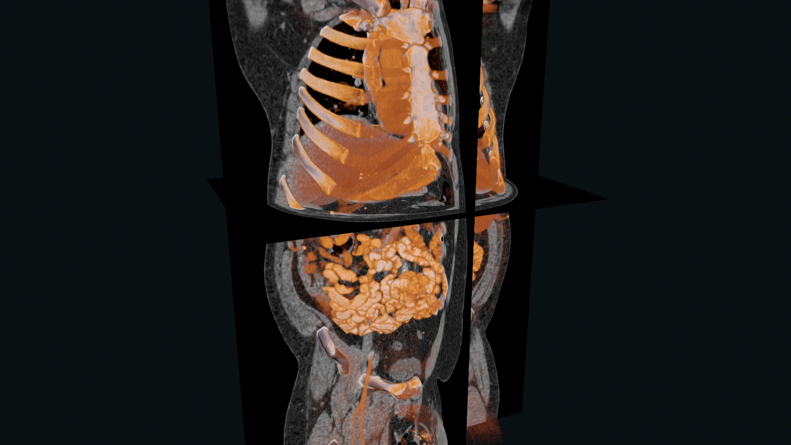
One study,many windows.
Lung, bone, soft tissue: one press, or drag it by hand.
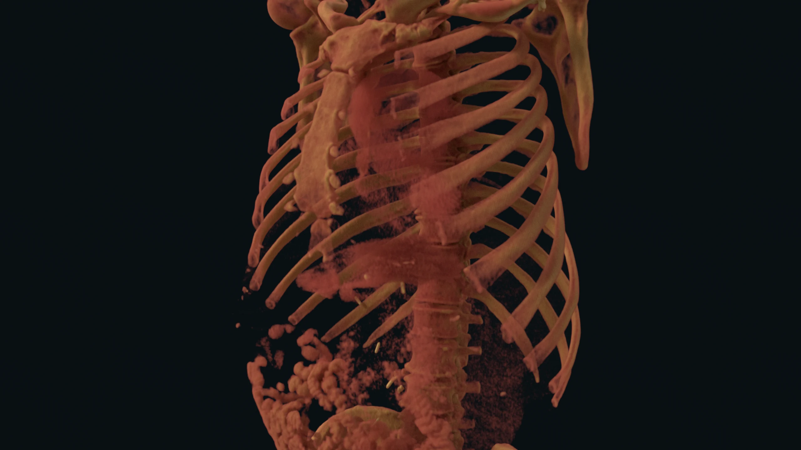
Volume renderingtransfer functions
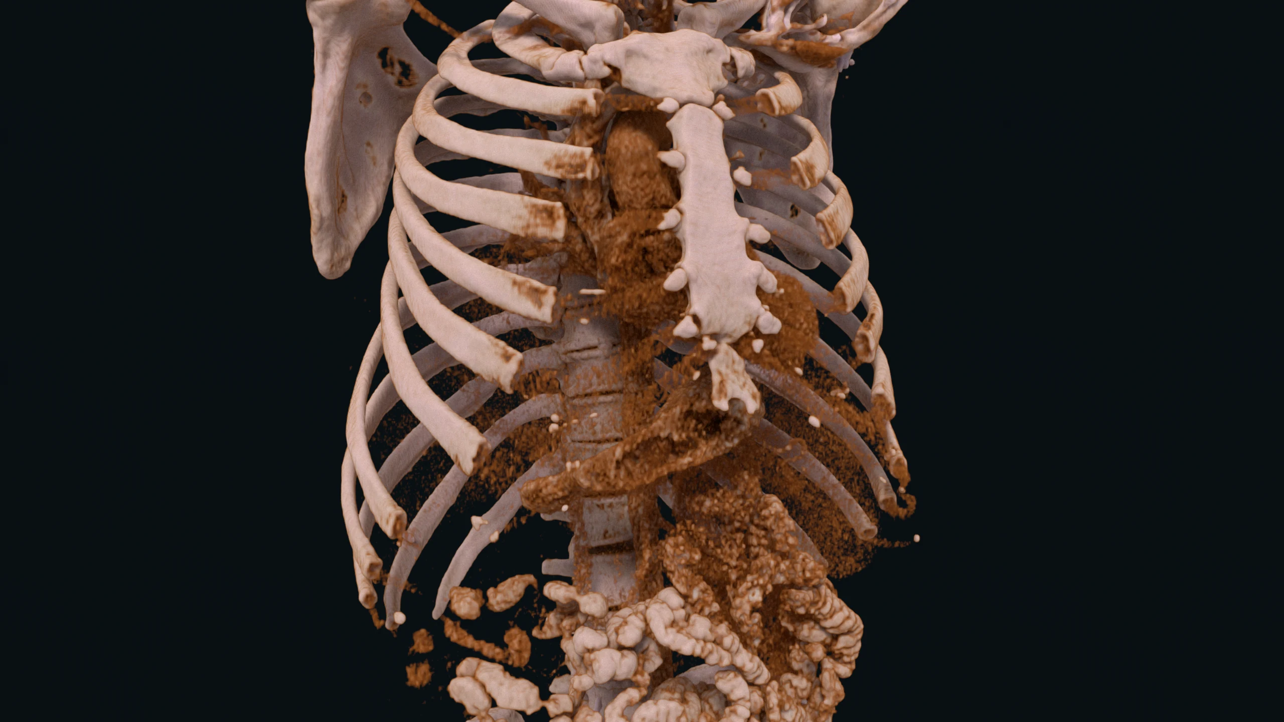
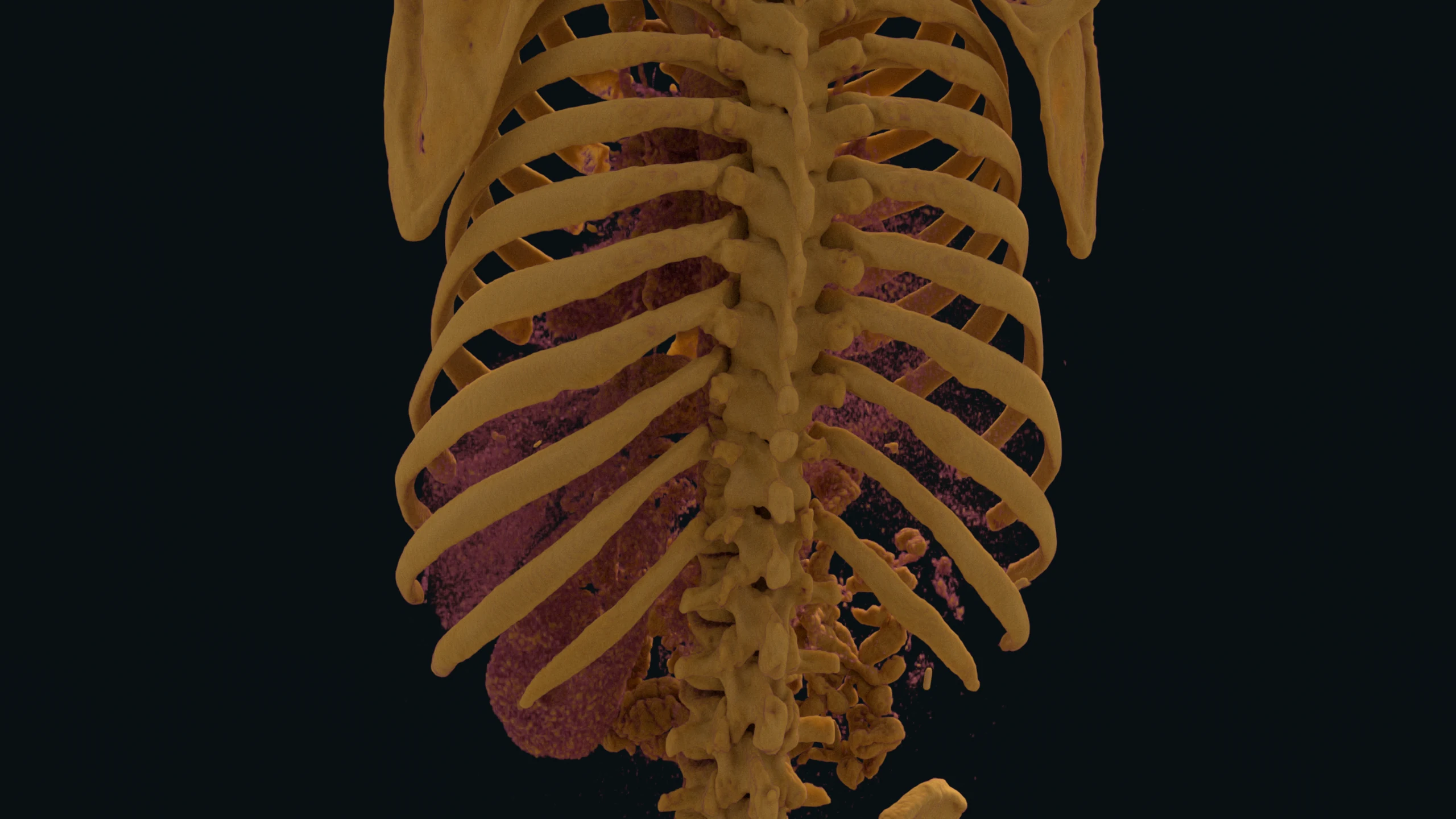
Light throughthe whole volume.
Pick a look, or draw your own curve. The light settles when you stop.
Measure and markon the slice
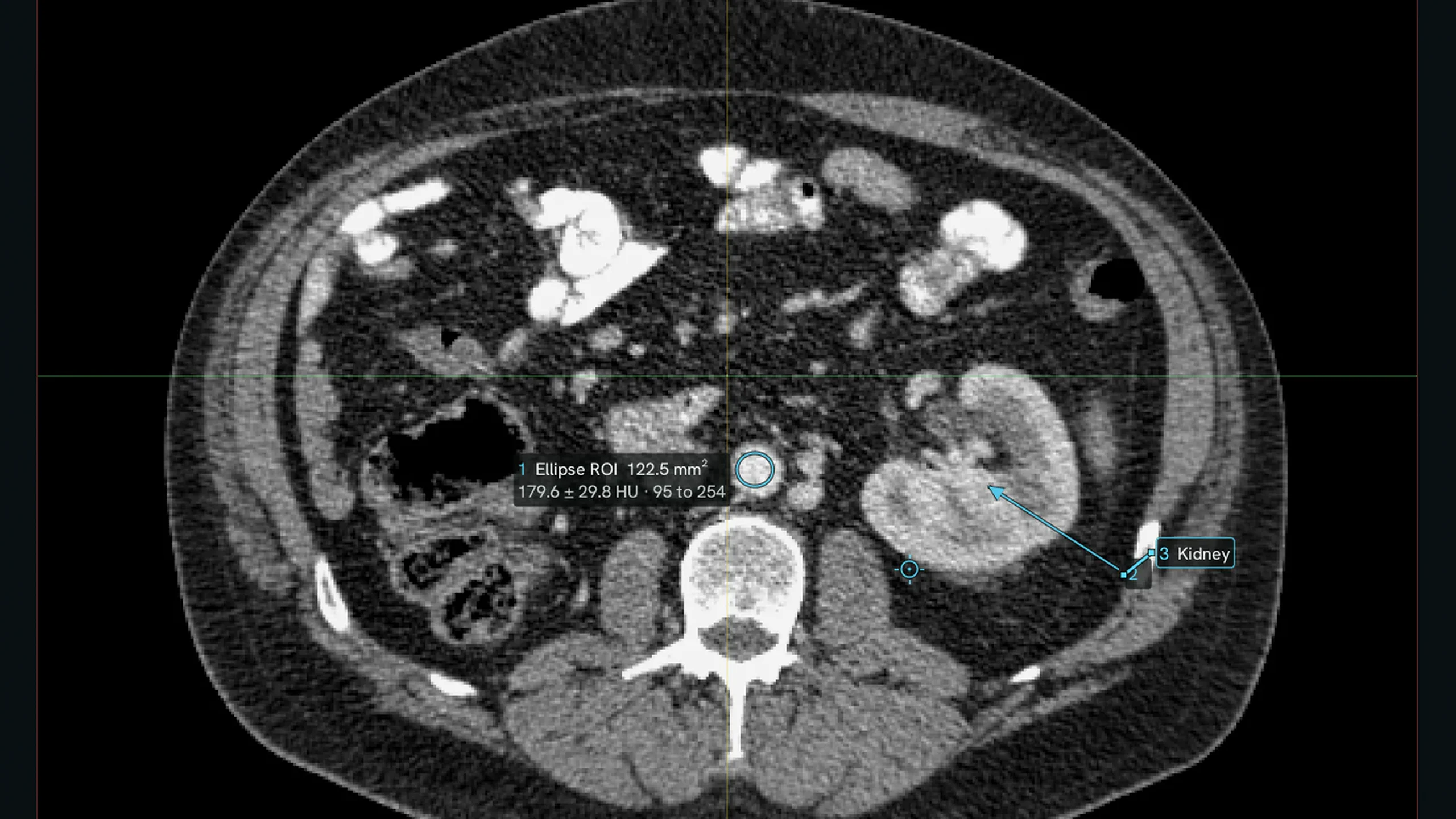
Draw on it.Keep the numbers.
- Text, arrows
- Ellipse, box, freehand regions with area and values
- Distances in millimetres
- Key images and a findings list as CSV or JSON
Inthe formats a scanner writes
Drop a folder.Get a volume.
It finds the series and the real geometry for you.
In 3Dstereo and headsets
A volume you canstand inside.
Stereo is built into the renderer. Volumes use it too.
Beyond medicineany stack of slices
Anything a scannercan see inside.
The same tools open industrial CT, micro-CT and fossils.
Parts and castings
Materials and micro-CT
Fossils
Heritage objects
Bring a study.See inside.
Part of the beta: try it on your own volumes.
For research, education and visualisation. Not a medical device; not for diagnosis.